Meet the Expert
Identification and history
Name: Zara
Report and medical history: dog, Jack Russell Terrier, Female neutered, 16y
No clinical signs, DUDE within normal limits.
Pot-belly, no PU/PD, increased liver enzymes on biochemistry.
Adrenal hyperadrenocorticism is suspected.
Diagnostics
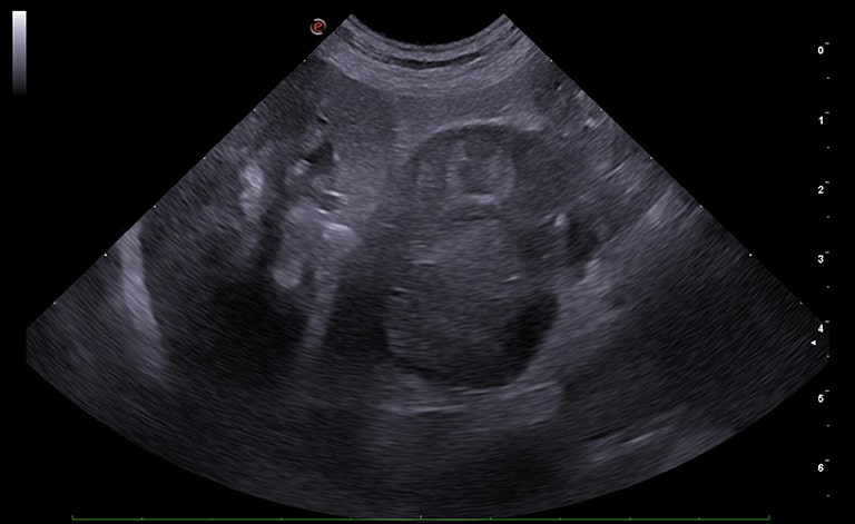
Right adrenal gland is markedly increased in size (3.3 cm) due to the presence of a heterogeneous mass. The latter is lining the caudal vena cava; however, a possible wall infiltration cannot be excluded.
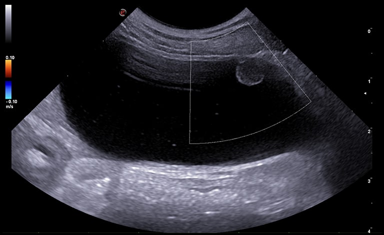
Urinary bladder presents hyperechoic non-gravity dependent material.
Wall thickness within normal limits.
An irregularly-rounded parietal lesion, protruding into the lumen and positive on color Doppler interrogation, is observed arising from the left ventro-lateral aspect of the urinary bladder neck.
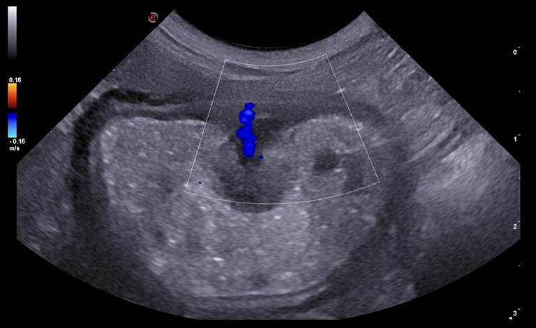
Presence of mixed fluid/gaseous content filling the gastric lumen.
A focal mucosal thickening of the greater curvature is observed, protruding into the lumen and positive to color Doppler interrogation.
Wall layering is preserved.
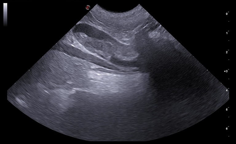
A fusiform endoluminal hyperechoic structure, partially obstructive, is observed in the aortic lumen between the renal artery and the aortic trifurcation.
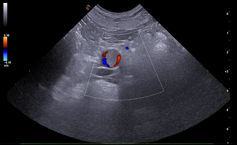
Transverse section of the aortic lesion previously described, with the hyperechoic partially obstructive endoluminal structure and surrounding blood flow.
Images were acquired with MyLab™9VET ultrasound system.
Conclusions and Treatment
Dr. Lapira Luca, DVM, Radiology Department OVU, University of Milan, Lodi, Italy
1. Right adrenal neoplasia (DDx adenoma, carcinoma, pheochromocytoma), likely secreting considering controlateral adrenal atrophy.
2. Non-specific bladder lesion DDx: polypoid cystitis vs neoplasia (e.g. TCC) vs idiopatic.
3. Gastric lesion DDx polyp vs neoplasia (e.g. lymphoma, carcinoma, leiomyoma).
4. Aortic thromboembolism.
Diagnostic confirmation of adrenal hyperadrenocorticism was obtained with dexamethasone suppression test; traumatic catheterization is proposed for further evaluation of bladder mass, and endoscopic biopsy for gastric lesion.
MyLab is a trademark of Esaote spa.
Product images are for illustrative purposes only. For further details, please contact your Esaote sales representative.
Technology and features are system/configuration dependent. Specifications subject to change without notice. Information might refer to products or modalities not yet approved in all countries.
Read other VET interviews
Canine ultrasonography
Report and medical history: Dog, Maltese, FS, 6 years old.
The patient requires examination for vomiting and diarrhea (hematochezia), presents with subicterus and temperature of 39.5°...
Canine echocardiography
Report and medical history: Dog, Corso, male, 3 months.
Echocardiographic evaluation requested by referring veterinarian for suspected congenital heart disease...
Equine echocardiography
Horse, thoroughbred Arab, male, 1 year old.
Grade 4 audible holosystolic murmur reported on both sides of chest, radiating ventrally to the right ...


