
Meet the Expert
Identification and history
Name: Amanda
Report and medical history: horse, Sella Italiano, female, 5 years old.
The subject shows tachycardia associated with reported Grade 3 holodiastolic murmur in the aortic area. In addition, the patient presents tachypnea, dicrotic inspiration and prolonged expiration.
Diagnostics
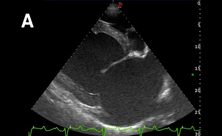
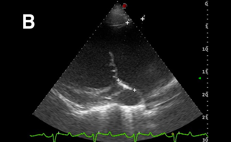
Right parasternal 4 chamber view (A) and left parasternal view of the left atrium and ventricle: significant dilation of the left atrium (v 166,4 mm; v.n 116 ± 7 mm) and the ventricle (systolic v 163.2 mm; v.n 112 ± 8 mm – diastolic v. 86.7) which present a globular aspect.
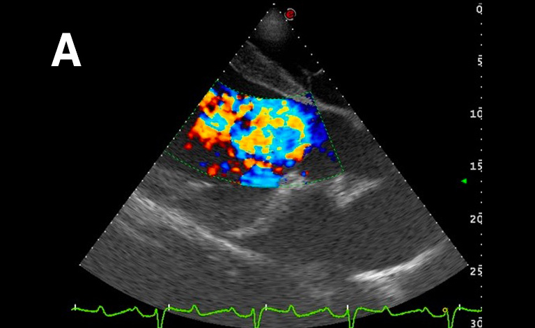
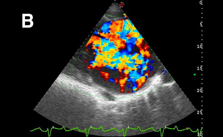
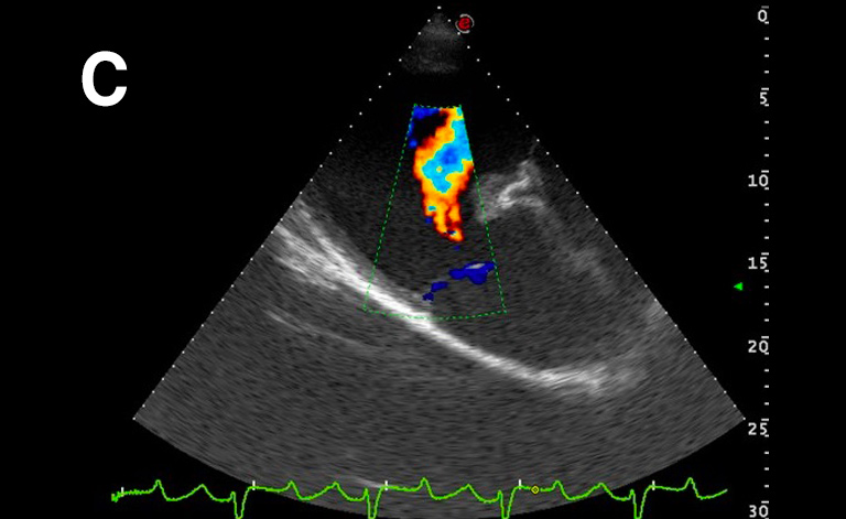
These images show: A) severe aortic regurgitation jet, B) severe mitral regurgitation jet; C) mild pulmonary regurgitation jet.
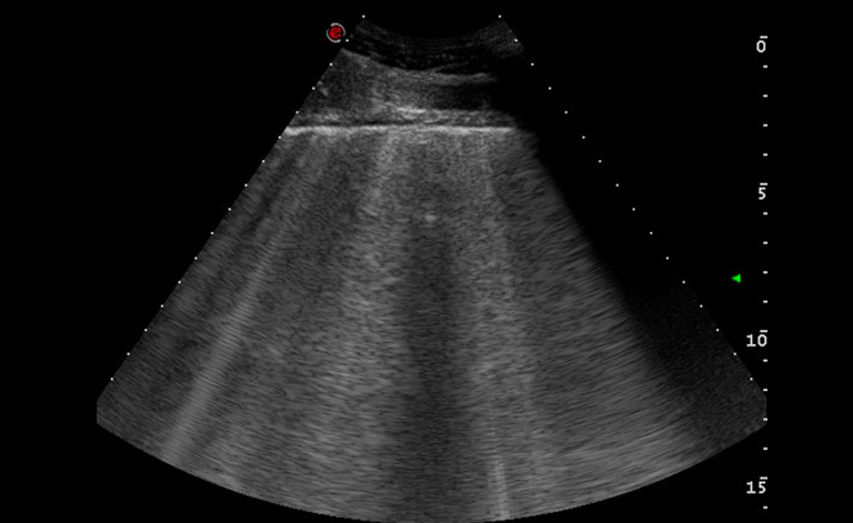
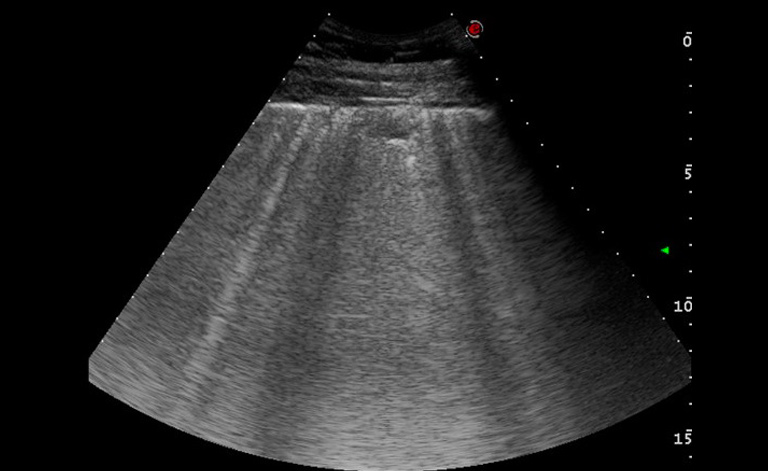
Auscultation of the chest found widespread wheezing, particularly intense in the apical region, bilateral.
The images show numerous comet tail artefacts widespread on both the right and left side that are suggestive of pulmonary edema.
Further exams: the subject also shows persistent sinus tachycardia (80 bpm) and systemic hypotension: the pressure measured by indirect non-invasive method was 70/89 mmHg. Arterial blood gas analysis found hypoxemia (pO2 80 mmHg).
Images were acquired with the MyLab™ClassCVET system.
Conclusions and treatment:
Equine Unit, University of Milan, University Veterinary Hospital of Lodi.
Left heart insufficiency associated with severe pulmonary edema and moderate systemic hypotension.
The patient has undergone symptomatic treatment with diuretics to reduce the pulmonary edema, and digoxin in order to improve the pumping action of the heart. The therapy has led to a progressive reduction of the pulmonary edema, improvement in pulmonary ventilation and a reduction in the baseline heart rate. The systemic pressure, closely monitored, has returned to the normal value range. In order to compensate for the possible loss of electrolytes caused by the diuretic therapy, it is advisable to monitor the level and, if necessary, administer supplements.
MyLab is a trademark of Esaote spa.
Product images are for illustrative purposes only. For further details, please contact your Esaote sales representative.
Technology and features are system/configuration dependent. Specifications subject to change without notice. Information might refer to products or modalities not yet approved in all countries.
Read other VET interviews

Canine echocardiography
Report and medical history: Dog, Corso, male, 3 months.
Echocardiographic evaluation requested by referring veterinarian for suspected congenital heart disease...

Canine ultrasonography
Report and medical history: Dog, Yorkshire Terrier, Female, 1 year old.
The patient presented with vomiting and diarrhoea while on heat. Haemorrhagic vomiting and diarrhoea persisted after sterilisation...

Canine ultrasonography
Dog, Jack Russell, neutered female, 11 years old.
Clinical visit requested for polyuria and polydipsia...