
Meet the Expert
Identification and history
Name: Pimpa
Report and medical history: dog, dachshund, female, 13 years old.
An abdominal ultrasound and echocardiogram was requested for loss of appetite, lethargy, and syncopal episodes.
Throughout the echocardiographic examination, a third-degree atrioventricular block is found on the basis of the electrocardiographic tracing.
Echocardiographic exam
Standard scan 2: In this video, the presence of a ventricular septal defect (VSD) is detected at the level of the membranous portion, near the right coronary aortic cusp, which indicates the presence of a systolic flow from the left ventricle to the right ventricle.
Visualization of the same defect is obtained in the short axis view from the right, at the level of the base of the heart.
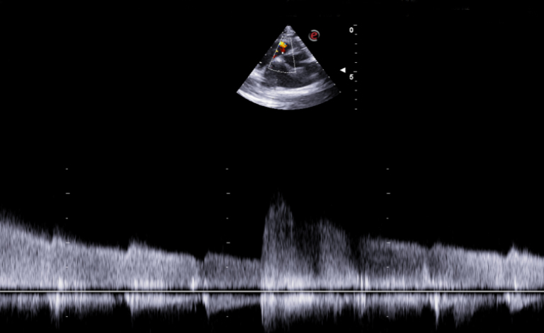
Systolic flow through the VSD, with left-to-right shunt, measured by continuous-wave Doppler; this flow appears to be poorly measurable because of the third-degree AV block.
Conclusions and treatment:

The echocardiographic diagnosis was perimembranous/supracristal ventricular septum defect.
Interventional procedure for correction of third-degree AV block by pacemaker implantation is recommended.
Echocardiographic images were acquired with the MyLab™X8VET system.
Claudio Bussadori, DVM, MD, PhD, Dipl. ECVIM (Cardiology)
Clinica Veterinaria Gran Sasso, Milan.
MyLab is a trademark of Esaote spa.
Product images are for illustrative purposes only. For further details, please contact your Esaote sales representative.
Technology and features are system/configuration dependent. Specifications subject to change without notice. Information might refer to products or modalities not yet approved in all countries.
Read other VET interviews

Feline Ultrasonography
Report and medical history: cat, Common European, spayed female, 16 years old.
The patient presented at our clinic with dejection, anorexia, hematochezia and vomiting. On clinical examination there was depressed sensory status ...

Canine Ultrasonography
Dog, Short-haired Dachshund, Female, 10 years old
Ultrasound checkup required for weight loss, PU/PD, recurrent vomiting, especially after ingestion of extra-diet foods ...

Canine musculoskeletal ultrasonography
Dog, Springer Spaniel, M, 4 years old
An ultrasound scan and orthopedic examination was requested for acute lameness in the front left leg...