Meet the Expert
Identification and history
Name: Cerry
Report and medical history: Dog, Miniature Poodle, FS, 5 YY.
The patient was taken to A&E due to poor appetite and despondency. On clinical examination, the dog presents an attitude of false kyphosis, sialorrhoea, and pain on palpation of the abdomen.
Diagnostics
At the level of the middle abdomen, colour Doppler examination indicated the presence of a parenchymatous, vascularised structure. The splenic parenchyma shows millimetric focal, hypo/isoechogenic lesions with blurred margins, not extending beyond the organ profile. The peritoneum appears diffusely hyperechogenic. Minimal layers of anechogenic effusion.
Hyperechogenic liver parenchyma without structural alterations. Distended gallbladder, severe parietal thickening, anechogenic endoluminal contents intermixed with finely corpusculated hyperechogenic material in suspension, not generating posterior shadow cone, also evident at the level of the common bile duct.
Mesenteric lymph nodes severely increased in size, hypoechogenic, with increased sphericity index.
Focal thickening of an intestinal loop, probably of jejunal relevance (7.2 mm), associated with loss of stratigraphic distinction.
Jejunal loop, mesenteric lymph nodes, peritoneal reactivity, layers of anecogenic effusion evidenced in the examined quadrants.
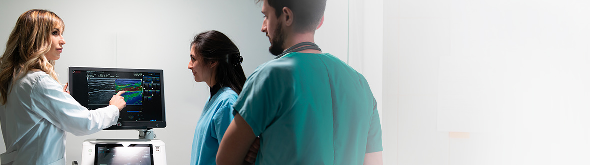
Images were acquired with MyLab™X90VET ultrasound system
Conclusions and Treatment
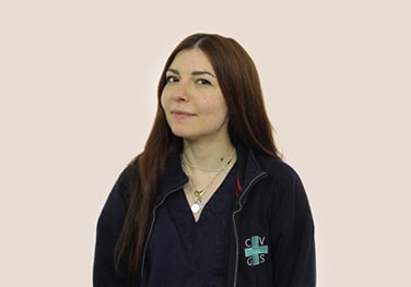
Signorelli Stefania, DVM, Clinica Veterinaria Gran Sasso, Milan, Italy
The ultrasound picture is compatible with an infiltrative process, probably of neoplastic origin (DD: Lymphoma, Carcinoma). Recommended cytological test for diagnostic purposes.

Canine ultrasonography
Report and medical history: Dog, Maltese, FS, 6 years old.
The patient requires examination for vomiting and diarrhea (hematochezia), presents with subicterus and temperature of 39.5°...

Canine ultrasonography
Report and medical history: dog, Jack Russell Terrier, Female neutered, 16y
No clinical signs, DUDE within normal limits. Pot-belly, no PU/PD, increased liver enzymes on biochemistry...

Canine ocular and abdominal Ultrasound
Report and medical history: Canine, half-breed, neutered male, 8 years old.
Differential diagnosis: suspected corneal ulcer complicated by bacterial infection...