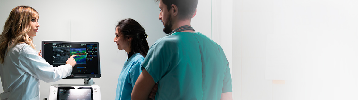
Meet the Expert
Identification and history
Name: Sissi
Report and medical history: Dog, Maltese, FS, 6 years old
The patient requires examination for vomiting and diarrhea (hematochezia), presents with subicterus and temperature of 39.5°. Silent past medical history, subject is in compliance with vaccine prophylaxis. No major problems to report in previous years. The patient lives mainly in an apartment and is fed pre-packaged food.
Diagnostics
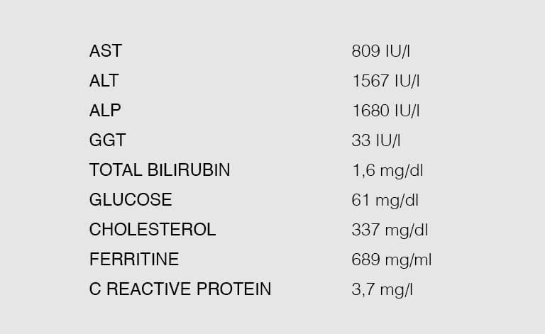
Stool examinations by flotation were performed and showed negative results. The Giardia test gave a negative result. The figure shows the result of the hematobiochemical examination. The hemogram is in the normal range with no major notes.
Ultrasound of the intestine
A complete abdominal ultrasound was performed that shows, among other findings, an abnormality of the small intestine.
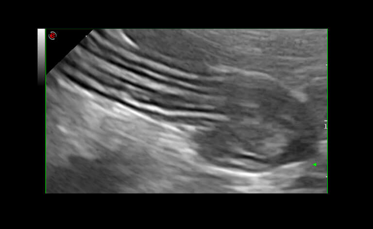
In the ultrasound image, the stratigraphy of the jejunal loop can be appreciated, which has an abnormality.
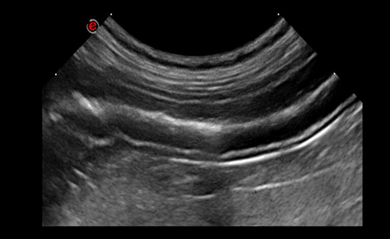
This ultrasound image shows the physiological stratigraphy of a jejunal loop, which its 5 layers.
Instead, from the ultrasound examination, in this subject appears a supernumerary layer. The hypotheses that are made as a justification for the supernumerary jejunal layers, in order of possibilities, are:
- infiltration/edema of the mucosa
- infiltration/edema of the submucosa
- hyper-echogenic band dividing the fasciae of the longitudinal muscles from the circular muscles
When the symptoms resolve, the finding disappears.
Ultrasound examination of the liver was then performed for the following reasons:
- the clinical symptoms lead us to suspect liver disease
- the clinical examination and its signs confirm possible alterations
- laboratory tests point towards a liver problem
- radiographic examination was not helpful.
Ultrasound of the liver
On ultrasound examination of the liver, an increased and nonhomogeneous echogenic pattern was found.
A liver biopsy was then performed.
Histological description:
Examined sections of 3 hepatic tru-cuts and 3 muscular/adipose tissue fragments. The hepatic histoarchitecture is disturbed due to an inflammatory and fibrotic process involving the portal spaces, with formation of fibrous porto-portal bridges and involvement of adjacent hepatic sheets. The infiltrate is characterized by a predominance of small Iymphocytes, fewer, mostly viable neutrophil granulocytes, macrophages phagocytizing cellular debris or brownish-yellow pigment (lipofuscin/hemosiderin), rarer plasma cells, and sporadic eosinophilic granulocytes. The neutrophilic infiltrate involves the adjacent hepatic sinusoids and multifocally the bile ducts, which are moderately hyperplastic. The hepatocytes near the portal spaces are characterized by severe hydropic-vacuolar degeneration and multifocal necrosis. Fibroplasia, fibrosis and neovascularization of the portal spaces, sporadic hematopoietic cells (megakaryoblasts), portal hemorrhages, and hepatocellular regenerative aspects (binucleation and karyomegaly) are evident.
Result of liver biopsy
The finding is related to severe neutrophilic, lymphocytic and necrotizing, chronic-active cholangiohepatitis with porto-portal fibrosis and hepatic regeneration. No morphologically detectable etiologic agents. Correlation to the overall clinicopathologic picture may be useful.
With liver ultrasound, portal vein flows were also assessed and splenic flow was found to be reversed (blue color coding).
A porto-systemic shunt was suspected, and to confirm this, a bubbling test was performed by inoculating a stirred mixture of blood and saline into the spleen. Simultaneously, an initial cardiac evaluation was performed.
The evaluation, concurrent with the bubbling test, showed small hyper-echogenic bubbles in the right atrium.
The patient was re-examined after about 10 days. The intestinal alterations have disappeared, the tests are improving but, despite the evidence of the shunt vessel the splenic flow has normalized.
Images were acquired with MyLab™Omega VET and MyLab™Eight VET ultrasound system.
Conclusions and Treatment

Dr. Giovanni Camali, DVM, Veterinary Clinic, Venice, Italy
What could the diagnosis be? Write and share your ideas with us.
Send us your hypotheses to: global.marketing@esaote.com
Read other VET interviews

Canine ultrasonography
Report and medical history: Dog, Miniature Poodle, FS, 5 YY.
The patient was taken to A&E due to poor appetite and despondency...

Canine ultrasonography
Report and medical history: dog, Jack Russell Terrier, Female neutered, 16y
No clinical signs, DUDE within normal limits. Pot-belly, no PU/PD, increased liver enzymes on biochemistry...

Canine ocular and abdominal Ultrasound
Report and medical history: Canine, half-breed, neutered male, 8 years old.
Differential diagnosis: suspected corneal ulcer complicated by bacterial infection...