
Meet the Expert
Identification and history
Name: horse, Arabian, male, 1 year old.
Report and medical history: Grade IV holosystolic murmur audible on both side of the chest, radiating ventrally and cranially on the right side.
Diagnostics
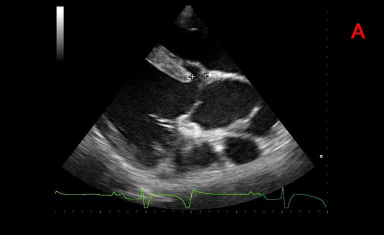
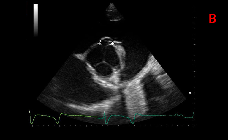
Right parasternal long axis view of the left ventricular outflow tract and aorta (A) and right parasternal short axis view at the level of the aortic valve (B): show a defect of the membranous portion of the interventricular septum located between the right coronary cusp of the aorta and the septal leaflet of the tricuspid valve. The maximum diameter of the defect, measured in the two mutually perpendicular planes, is 2.47 cm x 1.41 cm.
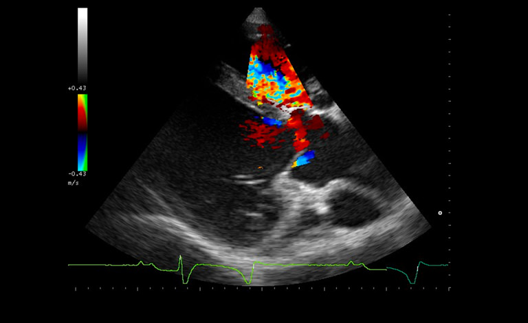
This image shows the systolic flow shunt from the left to the right ventricle by means of Color flow Doppler. The maximum velocity of the left-right transventricular flow measured by continuous Doppler is 4.82 m/s.
Images were acquired with the MyLab&reade;5VET system.
Conclusions and treatment:

Equine Medicine Unit, University of Milan, University Veterinary Hospital of Lodi.
Membranous ventricular septal defect at the level of the right coronary cusp of the aortic valve.
This is a congenital heart defect disease. Given the localization in the membranous portion, the dimension below 2,5 cm in both orthogonal planes and the shunt velocity greater than 4m/s, the prognosis is good and the
horse can have a successful athletic career.
MyLab is a trademark of Esaote spa.
Product images are for illustrative purposes only. For further details, please contact your Esaote sales representative.
Technology and features are system/configuration dependent. Specifications subject to change without notice. Information might refer to products or modalities not yet approved in all countries.
Read other VET interviews

Canine ultrasonography
Dog, Jack Russell, neutered female, 11 years old.
Clinical visit requested for polyuria and polydipsia...

Canine Echocardiography
Report and medical history: dog, dachshund, female, 13 years old.
An abdominal ultrasound and echocardiogram was requested for loss of appetite, lethargy, and syncopal episodes ...

Canine musculoskeletal ultrasonography
Dog, Springer Spaniel, M, 4 years old
An ultrasound scan and orthopedic examination was requested for acute lameness in the front left leg...