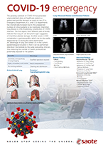
The growing outbreak of COVID-19 has generated unprecedented stress on healthcare systems at global level and the demand of Point-of-Care US in Emergency, ICU and CCU departments dramatically increased due to unexpected number of critical patients to be monitored. Interstitial lung disease is a life-threatening complications of COVID infection.
The first reports from different parts of world indicate that lung US can document signs suggestive for interstitial-alveolar damage. Another severe COVID complication is perimyocarditis, that can be also easily diagnose by US during the same exam.
Advantage of lung and cardiac US in a current epidemiologic situation is that it can be performed directly at bed-side by the same evaluating clinician, thus reducing the number of health workers potentially exposed to the patient.
Benefits
- Sensitivity and specificity in lung-imaging
- Agile, immediate and mobile
- No ionizing radiation
Warnings
- Qualified operators required
- Results interpretation
- Cleaning and disinfection
Lung Ultrasound Normal and Abnormal Patterns

A-lines

Regular pleural line, B-lines

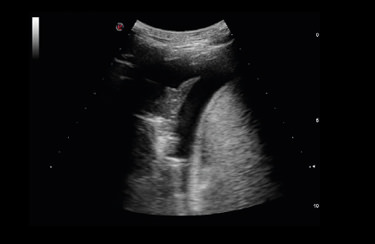
Large pleural effusion, lung consolidation

Irregular pleural line, B-lines
Normal findings
“SPAA”- Lung Sliding
- Lung Pulse
- A-Lines
- Short Vertical Artifacts
Abnormal findings
Pneumothorax “3A-2P”- Absence of Lung Sliding
- Absence of Lung Pulse
- Absence of Comete Tail/Vertical Artifacts
- Presence of Lung Point
- Presence of A-Lines
- Absence of A-Lines
- B-Lines
- Consolidation
- Negative Curtain Sign
- Positive Spine Sign
- Presence of Fluid

Complete
- High performance in all the applications
- Large probes portfolio, convex, linear and phased-array
- Advanced configurations, including TE probe or Strain package
Fast
- 2 probes connectors Up to 4 with multiconnector
- Touchscreen with intuitive menus
- Long-duration battery and quick boot-up time (15”)
Compact
- Full screen mode
- Swivelling monitor
- Agile trolley easy with 4 swivelling wheels
Connected
- Follow-up & Multi-modality options to retrieve other imaging modalities
- eStreaming for real-time imaging streaming
- eTablet & MyLab™Remote for remote storage and control

Low-frequency probe to scan lung parenchyma and commonly used in emergency for abdominal organs.

High-frequency probe to scan pleural area and superficial structures. Commonly used to scan vessels and support lines’ placement.

Low-frequency phased-array probe to scan the lung and commonly used for heart functionality monitoring.
Scientific references
- Proposal for international standardization of the use of lung ultra-sound for COVID-19 patients; a simple, quantitative, reproducible method.
Soldati G, Smargiassi A, Inchingolo R, Buonsenso D, Perrone T, Briganti DF, Perlini S., Torri E., Mariani A., Mossolani E.E., Tursi F., Mento F., Demi L. - J Ultrasound Med. 2020 Mar 30. doi: 10.1002/jum.15285 -
Is there a role for lung ultrasound during the COVID-19 pandemic?
Soldati G., Smargiassi A., Inchingolo R., Buonsenso D., Perrone T., Briganti D.F., Perlini S., Torri E., Mariani A., Mossolani E.E., Tursi F., Mento F., Demi L. - J Ultrasound Med. 2020 Mar 20. doi: 10.1002/jum.15284. -
Point-of-Care Lung Ultrasound findings in novel coronavirus disease-19 pneumoniae: a case report and potential applications dur-ing COVID-19 outbreak
Buonsenso D., Piano A., Raffaelli F., Bonadia N., De Gaetano Donati K., Franceschi F. - Eur. Rev. Med. Pharmacol. Sci. 2020 Mar; 24(5):2776-2780. doi: 10.26355/eurrev_202003_20549.
-
Accuracy of Lung Ultrasound in Patients with Acute Dyspnea: The In-fluence of Age, Multimorbidity and Cognitive and Motor Impairment
Vizioli L., Forti P., Bartoli E., Giovagnoli M., Recinella G., Bernucci D., Masetti M., Martino E., Pirazzoli G.L., Zoli M., Bianchi G. - Ultrasound in Medicine & Biology, Volume 43, Issue 9, September 2017, Pages 1846-1852, ISSN 0301-5629 -
Role of lung ultrasound in paediatric intensive care units: comparison with bedside chest radiography
Mughetti M., Napoli G., Chiesa A.M., Ciccarese F., Bertaccini P., Zompatori M. - European Congress of Radiology 2017 - Poster No.: B- 0161-DOI:0.1594/ecr2017/B-0161 - dx.doi.org/10.1594/ecr2017/B-0161 -
Influence of positive end-expiratory pressure on myocardial strain assessed by speckle tracking echocardiography in mechanically ven-tilated patients.
Franchi F., Faltoni A., Cameli M., Muzzi L., Lisi M., Cubattoli L., Cecchini S., Mondillo S., Biagioli B., Taccone F.S., Scolletta S. - Biomed. Res. Int. 2013;2013:918548. doi: 10.1155/2013/918548. Epub 2013 Aug 28. -
Early recognition of the 2009 pandemic influenza A (H1N1) pneu-monia by chest ultrasound
Testa A., Soldati G., Copetti R., Giannuzzi R., Portale G., Gentiloni-Silveri N. - Critical Care 2012, 16 (1), R30, 1012 Febr. 17 http://ccforum.com/content/16/1/R30
Related articles
Critical Care examinations made easier during the Covid-19 era
Never stop striving for easy Scanning and easy Quantification in Critical Care in Cardiology, Abdominal, Lung, Vascular, TCD.
Lung Ultrasound in COVID Patients
COVID-19 pneumonia is characterized by alveolar edema with prominent proteinaceous exudates, vascular congestion, patchy inflammatory clusters with fibrinoid material, alveolar epithelial hyperplasia, and fibroblastic proliferation...
Interview to Esaote CEO: “We’ll answer the call of Europe and the USA too.”
First, Esaote came to the aid of China, a country with which the company has strong connections, given that their shares are owned bya group of six Chinese industrial (Wandong, Yuwell, Kangda) and financial companies (Yunfeng, Shanghai Ftz, Tianyi).
Esaote assembles and delivers 103 portable ultrasound scanners in just 3 days
Esaote won a tender launched by Consip on behalf of Civil Protection for the distribution of diagnostic equipment in Italy to face COVID-19 emergency.
The eyes above the masks. Esaote field service engineers
We’ve asked six of them to observe the situation on the front line and share the ways in which their work has changed.
What we can do
We have a duty to develop a complete understanding of the limits within which we can carry out our production activities.
Probes and Agents
The table here included list MyLab ultrasound probes and the recommended cleaning, disinfection and sterilization agents and systems
Covid Webinar - Lung Ultrasound for the Covid-19 era
Covid Webinar - Lung Ultrasound for the Covid-19 era (F. Piscaglia, F. Stefanini, V. Salvatore)
Interview with Dr. Vito Cianci
The emergency room is the front office of the hospital. The doctors who work there are used to making quick decisions: what has changed with the Covid-19 emergency? What are the potentials and limitations of lung ultrasound?
Interview with Dr. Giovanni Battista Fonsi
In the daily management of the COVID-19 emergency, was it necessary for health professionals to review their way of doing ultrasound? How important is it to be able to carry out assessments on multiple districts, if necessary, through ultrasound?


